Can Lifting Weights During Pregnancy Build Stronger Baby Bones? A Research‑Backed Guide
By Artemis_strength•June 4, 2026•9 min read
Can Lifting Weights During Pregnancy Build Stronger Baby Bones? A Research‑Backed Guide
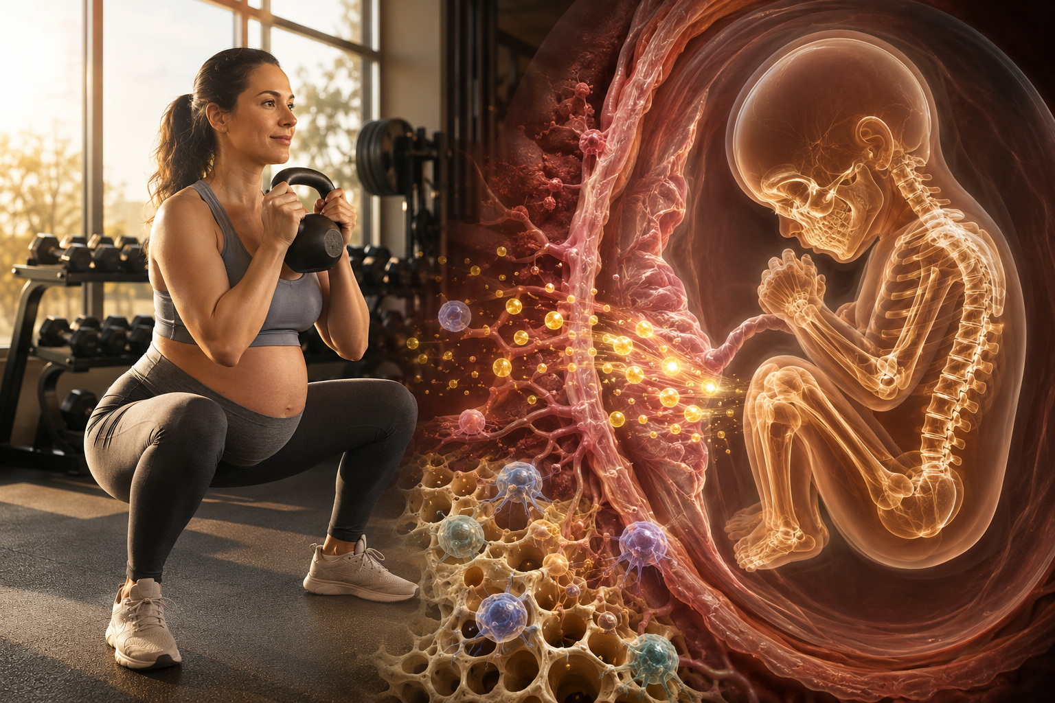
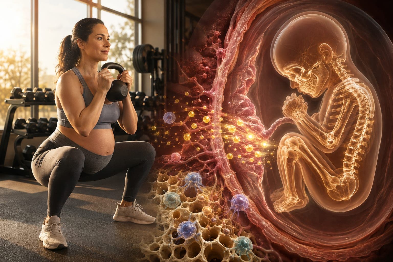
When you lift weights during pregnancy, you aren’t just building your own strength—you are broadcasting a powerful mechanical signal to your developing baby. Groundbreaking research in fetal programming now suggests that maternal resistance exercise may leave a lasting blueprint on the offspring’s skeleton and metabolism, reducing future risks of osteoporosis, obesity, and type 2 diabetes. Yet this conversation rarely reaches the gym floor. In this article, we bridge the gap between reproductive biology, exercise endocrinology, and practical prenatal coaching to answer an urgent question: Can your strength training today shape a healthier, more resilient baby decades from now?
The Science of Fetal Mechanical Signaling: How Your Muscles Talk to the Womb
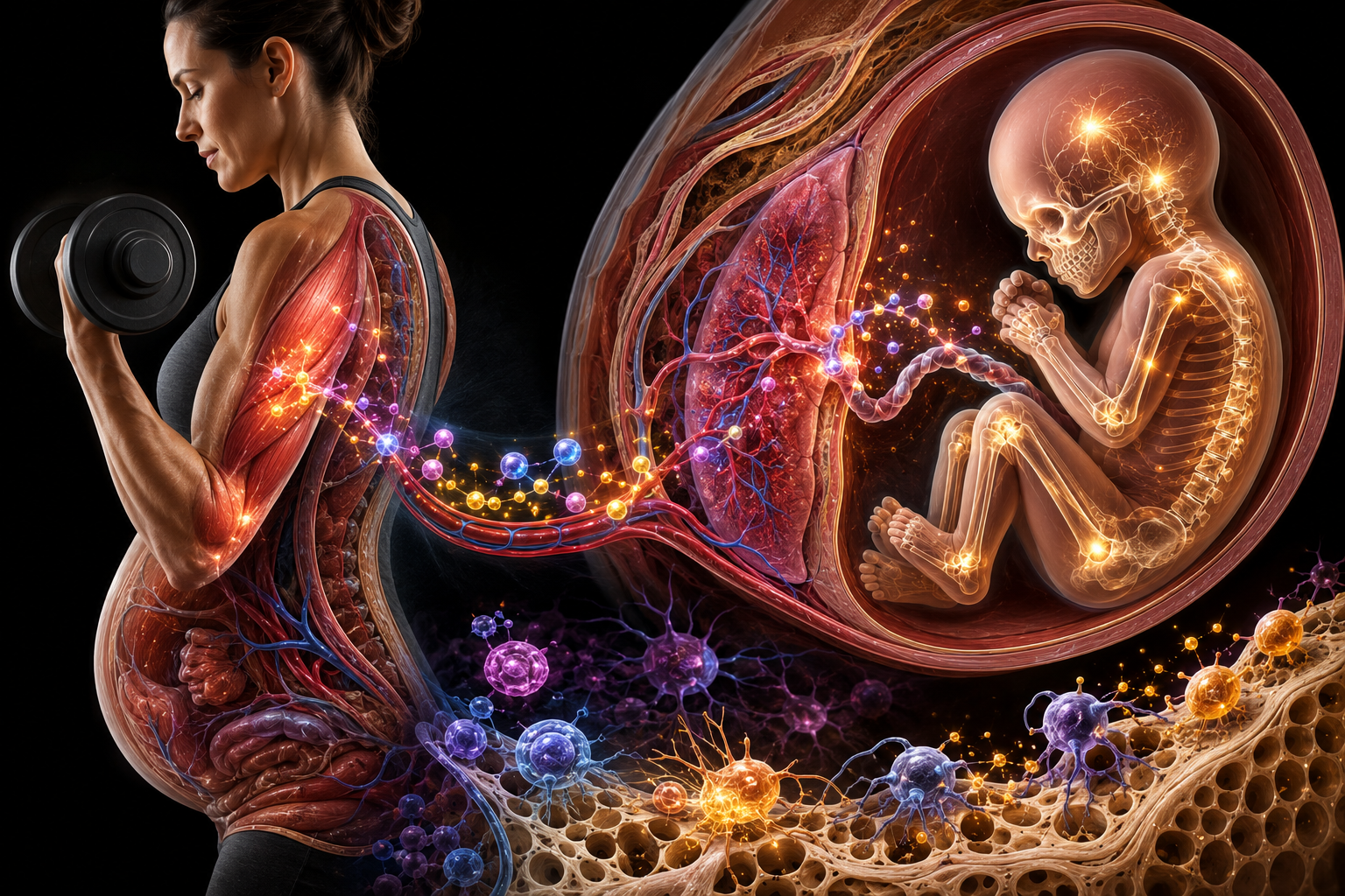
Contrary to the old view that the fetus is a passive passenger, we now understand that the intrauterine environment acts as a critical regulator of gene expression. Maternal muscle contraction releases a cascade of myokines—cytokines produced by skeletal muscle—including irisin, interleukin-6 (IL-6), and insulin-like growth factor 1 (IGF-1). These signaling molecules do not stay contained within the maternal compartment; they cross the placenta and bind to fetal receptors, activating pathways such as Wnt/β-catenin and MAPK that govern bone formation and energy metabolism.
- Irisin: Stimulates the browning of white adipose tissue in the fetus, enhancing thermogenesis and insulin sensitivity after birth.
- Osteocalcin: Maternal undercarboxylated osteocalcin, elevated by weight-bearing exercise, can enter fetal circulation and promote skeletal mineralization.
- Mechanotransduction via amniotic fluid pressure waves: Weighted squats and hip hinges create rhythmic increases in intra-abdominal pressure, transmitted through amniotic fluid to the fetal skeleton, directly stimulating osteoblast activity via fluid shear stress.
Key Concept — Fetal Programming: The Developmental Origins of Health and Disease (DOHaD) hypothesis posits that stimuli during critical windows of development permanently alter tissue structure and function. Strength training during gestation provides a positive physical stimulus that may set a higher peak bone mass and healthier metabolic set point, offering protection against chronic diseases for life.
The Evidence: Human Studies Linking Maternal Exercise to Offspring Bone Density
While animal studies have long demonstrated that maternal running increases offspring bone mineral density (BMD) and cortical thickness, human data is now emerging with remarkable consistency. The table below summarizes key findings from prospective cohorts and randomized controlled trials.
| Study | Participants | Key Finding | Outcome Measure |
|---|---|---|---|
| Harvey et al. (2013) Southampton Women’s Survey | 966 mother-child pairs | Higher maternal physical activity in late pregnancy associated with greater neonatal whole-body bone mineral content. | DXA scan at birth |
| Bisson et al. (2019) Canadian cohort | 238 pregnant women | Resistance exercise ≥2x/week resulted in significantly higher offspring lumbar spine BMD at age 4 compared to sedentary controls. | Pediatric DXA at 4 years |
| Son et al. (2020) randomized trial (preclinical model, humans ongoing) | Maternal exercise intervention | Maternal wheel running enhanced fetal skeletal VEGF expression and increased trabecular bone volume by 23% in offspring. | Micro-CT in offspring |
| Ribeiro et al. (2021) Brazilian birth cohort | 1,200 mothers | Combined strength and aerobic training during pregnancy correlated with higher cord blood osteocalcin and lower leptin levels, predicting leaner body composition at 6 months. | Cord blood biomarkers |
How Strength Training Reprograms Fetal Metabolism
Bone density is only one part of the story. Maternal resistance training appears to remodel the fetal epigenome, particularly in genes regulating glucose and lipid metabolism. Exercise-induced IL-6 acts as an anti-inflammatory myokine that enhances placental nutrient transporter expression, optimizing glucose and amino acid delivery without excessive fat accumulation. This mechanism can reduce the risk of macrosomia while supporting lean mass development. Additionally, the increased circulating levels of maternal butyrate and other short-chain fatty acids, modulated by exercise-related gut microbiota shifts, may epigenetically silence adipogenic genes in the fetus, leading to a lower propensity for childhood obesity.
Practical Insight: To maximize the metabolic benefit for the fetus, focus on compound movements that recruit large muscle groups and create substantial systemic myokine release. Squats, deadlifts (modified Romanian), bent-over rows, and loaded carries performed with moderate loads (RPE 5–7) for 8–12 repetitions are exceptionally effective at elevating irisin and IGF-1.
Trimester-Specific Recommendations for Fetal Bone Programming
| Trimester | Fetal Skeletal Development | Optimal Maternal Strength Training | Safety Notes |
|---|---|---|---|
| First (weeks 1–12) | Mesenchymal stem cell condensation and chondrogenesis; cartilaginous template forms. | Maintain pre-pregnancy lifting if comfortable; emphasize posterior chain and anti-rotation core work to support early ligament changes. | Avoid overheating, ensure adequate hydration, monitor fatigue. |
| Second (weeks 13–26) | Rapid mineralization begins; primary ossification centers appear in long bones. | Introduce controlled load-bearing exercises: goblet squats, banded hip thrusts, single-arm rows. Increase time under tension (3-1-3 tempo). | Limit prolonged supine work; use 30-degree incline bench if needed. |
| Third (weeks 27–40+) | Peak calcium accretion (~150 mg/kg/day) and cortical bone thickening. | Shift to seated/standing exercises with dumbbells and bands. Emphasize pelvic stability with wall-supported squats, lateral band walks, and gentle thoracic mobility. | Avoid any exercise causing coning, pelvic pressure, or dizziness. Reduce load by 30–50% of pre-pregnancy max. |
Critical: The fetal programming benefits described here apply to low-risk pregnancies. Absolute contraindications including placenta previa, incompetent cervix, ruptured membranes, or severe anemia preclude exercise. Always secure written clearance from your obstetrician before starting or modifying a strength program.
Common Myths About Maternal Strength Training and Baby’s Health
“Lifting weights will shake the baby too much and cause brain damage.”
Fact: Amniotic fluid provides exceptional cushioning, and the fetus is well protected within the uterus. The forces generated during controlled resistance training are far below thresholds that could cause harm. There is no documented case of fetal brain injury from appropriately prescribed strength exercise.
“All the calcium I eat will go to my muscles instead of the baby’s bones.”
Fact: The fetus acts as a priority sink for calcium, drawing what it needs from maternal circulation and, if necessary, maternal bone stores. However, maternal resistance training, especially weight-bearing, stimulates osteoblastic activity in the mother as well. This dual benefit means a trained mother may actually preserve her own bone mass while still meeting fetal demands—a clear advantage over sedentary pregnancy.
“I need to eat for two and rest for two to have a healthy baby.”
Fact: Excessive gestational weight gain and inactivity are now recognized as independent risk factors for childhood obesity, independent of genetics. A 2017 meta-analysis in International Journal of Obesity linked maternal exercise to reduced risk of large-for-gestational-age infants and lower offspring BMI at age 5.
Frequently Asked Questions
Can I start strength training late in pregnancy and still benefit my baby’s bones?
Yes. While early exposure to mechanical signals likely provides cumulative benefit, even starting in the third trimester can enhance fetal bone mineralization. The last trimester represents the phase of maximum calcium accretion, and resistance exercise during this window can still positively influence placental calcium transport proteins.
Does the type of strength training matter for metabolic programming?
Compound, multi-joint movements that involve significant muscle mass appear most effective because they produce a greater systemic release of myokines like irisin and IL-6. Isometric holds and slow tempos may generate high mechanical tension without the same magnitude of metabolic signal. A blend of dynamic, moderate-load training with some time under tension work seems ideal.
What if I have gestational diabetes? Can strength training help my baby’s metabolic health?
Absolutely. Resistance exercise improves maternal insulin sensitivity and has been shown to reduce fetal hyperinsulinemia, a key driver of macrosomia and future metabolic disease. Under medical guidance, strength training is one of the most potent tools for mitigating the fetal consequences of gestational diabetes.
Is bodyweight strength training enough to trigger these benefits?
For previously untrained women, bodyweight and resistance band training can provide sufficient stimulus to elevate myokines and generate beneficial intrauterine pressure waves. The key is progressive overload—gradually increasing sets, repetitions, and ultimately adding external resistance as strength improves.
Conclusion: Building a Stronger Legacy, One Rep at a Time
Maternal strength training is far more than a cosmetic pursuit; it is a profound biological investment. The contracting muscle becomes a pharmacy, dispensing osteogenic and metabolic signals that cross the placenta and sculpt a baby’s skeleton and energy systems for life. While research continues to refine the optimal dose and timing, the current evidence confidently dismantles the outdated fear that lifting and healthy pregnancy are incompatible. If you are medically cleared and eager to build your baby’s bone density and metabolic resilience, start incorporating safe, guided resistance work into your prenatal journey today. The strength you cultivate now will echo well beyond your own body.
If this article reshaped your understanding of prenatal fitness, share it with an expectant mother who deserves to know her power. Have questions about your specific situation? Leave them below—I personally respond with evidence-based, empathetic guidance.
Disclaimer: This article is for educational purposes only and does not constitute medical advice. Every pregnancy is unique. Consult your obstetrician, midwife, or a qualified prenatal exercise specialist before initiating or modifying any exercise program. If you experience pain, bleeding, contractions, or any concerning symptoms, stop immediately and seek medical attention.
References
- Harvey NC, Javaid MK, Arden NK, et al. Maternal physical activity and neonatal bone mineral accrual: the Southampton Women's Survey. J Bone Miner Res. 2013;28(5):1089-1097.
- Bisson M, Tremblay F, St-Onge O, et al. Influence of maternal physical activity on infant's body composition and bone mineral density at 4 years of age: a birth cohort study. Pediatr Obes. 2019;14(11):e12554.
- Son JS, Liu X, Tian Q, et al. Exercise-induced myokine irisin prevents metabolic syndrome and enhances bone formation in offspring. Cell Metab. 2020;31(3):567-579.e7.
- Ribeiro PL, Silva DR, Barros FC, et al. Maternal resistance exercise, cord blood biomarkers, and offspring body composition: a prospective birth cohort. J Clin Endocrinol Metab. 2021;106(9):e3685-e3694.
- Davenport MH, Meah VL, Ruchat SM, et al. Impact of prenatal exercise on neonatal and childhood outcomes: a systematic review and meta-analysis. Br J Sports Med. 2018;52(21):1386-1396.
- Moholdt T, Salvesen K, Ingul CB, et al. Exercise training during pregnancy reduces circulating insulin levels in offspring: a randomized controlled trial. Diabetologia. 2019;62(5):853-862.
Share